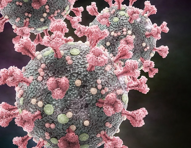
In a recent preprinted paper on bioRxiv, US researchers demonstrate the central role of the insertion of the furine excision site into the complex glucoprotein of coronavirus 2 (SARS-CoV-2) of severe acute respiratory syndrome plays in viral replication and pathogenesis, highlighting, in turn, that they want to monitor mutations the progression of treatments and vaccines.
While coronavirus disease (COVID-19) continues to have an effect on the world, a sufficiently good understanding of the SARS-CoV-2 replication and virulence houses would likely reveal possible avenues to stop the disease. So far, most of the attention has been directed at complex glucoprotein responsible for binding to the receptor and moving input.
In short, after the popularity of the angiotensin 2 conversion enzyme receptor (ACE2), complex glycoprotein is divided into two s (S1/S2 and S2′) to facilitate viral access to the cell. As a result, many researchers have focused their attention on a potentially critical insertion of a discovered furine excision upstream of S1 excision into complex glucoprotein.
Related stearal excision sites have been observed in other virulent pathogens such as avian influenza, human immunodeficiency virus (HIV) and Ebola. More importantly, furin excision sites can be discovered in a large number of other members of the coronavirus circle of relatives, adding MERS-CoV. Therefore, the acquisition of the furin excision site can be observed as a “function gain”, which in all likelihood opens the door to the virus to jump to humans.
The applicable question is whether the furine cleavage site plays a significant role in the infectiousness and pathogenesis of SARS-CoV-2. This challenge was recently addressed through an organization of researchers from the medical branch of the University of Texas, Emory University, Icahn Medical School in Mount Sinai, the University of Texas Health Science Center in Houston, and Bowie State University in the United States. States.
In this manuscript, scientists necessarily used an opposite genetic formula to generate a SARS-CoV-2 mutant without the insertion of the furina division site. Specifically, a reverse genetic formula for SARS-CoV-2 WA1 isolate (originally received from the US CDC), spawned a mutant virus that suppressed the insertion of 4 amino acids (PRRA).
Vero E6 mobile phones (derived from African green monkey renal epithelial mobiles) or Calu3 mobiles (derived from a mobile line of non-small mobile lung cancer with epithelial morphology and adhering growth) were inflamed with wild or mutant viruses. Several strategies tested the relative degrees of viral proteins.
The researchers then assessed the ability of the PRRA mutant in relation to wild-type SARS-CoV-2 in a competitive test. Specifically, using plaque-forming sets to enter, they combined wild-type and mutant viruses in other proportions into Vero E6 cells, and then evaluated their overall aptitude with an oppos transcription PCR approach.
For in vivo studies, hamsters faced wild or mutant viruses through intranasal inoculation and were then observed the progression of clinical disease. The review also included a wide variety of complex genetic methods, such as deep sequencing analysis, phylogenetic tree structure, use of thermal series identity maps, and structural modeling.
In short, the loss of the furine excision site at the SARS-CoV-2 peak had a significant effect on infection and pathogenesis. The mutant-PRRA had improved replication houses and greater skill in Vero E6 cells compared to wild SARS-CoV-2, as well as a reduced complex remedy. In contrast, the mutant-PRRA dims in Calu3 cells and had a modified complex glucoprotein remedy compared to Vero cells.
In addition, plaque relief neutralization tests performed with COVID-19 patient serums and monoclonal antibodies opposed to the complex glucoprotein receptor binding domain uncovered a change, the mutant virus caused constant relief in neutralization titers.
In hamsters, furine site loss alleviates SARS-CoV-2-induced disease but does not suppress replication of the PRRA virus. However, despite a mild illness, the first infection, hamsters inflamed with ‘PRRA’ were in fact protected from further provocation with wild-type SARS-CoV-2, indicating the generation of a physically powerful immune response.
“In general, the knowledge presented in this manuscript illustrates the critical role of inserting furin excision into the protein complex playing in SARS-CoV-2 infection and pathogenesis,” the review authors verify their findings in the bioRxiv preprinted document.
Biologically speaking, the loss of the furine site displaces the remedy of complex glycoprotein in a way that depends on the type of mobile. In addition, the mutant PRRA virus is impaired in its ability to reflect certain types of mobile and cause in vivo diseases. However, the effects are confusing due to the increased replication and skill observed on Vero mobiles.
Finally, the changed antibody neutralization profiles reveal a critical desire to examine this mutation in researching the characteristics of the SARS-CoV-2 remedy and long-term vaccine applicants. In addition, understanding the main points of the furin split site can even help us avoid potential long-term pandemics caused by coronavirus.
bioRxiv publishes initial clinical reports that are not peer-reviewed and therefore should not be considered conclusive, the consultant’s clinical practice/health-related behaviors, nor treated as established information.
Highlights: – mutant dPRRA attenuated in vitro and in vivo – The mutant has a fitness merit in Vero E6 versus WT – PRNT50 Mutant Change Values ‘SARSCoV2’ COVID19
Written by
Dr. Tomislav Me-trovio is a physician (MD) with a physicist. in biomedical and fitness sciences, specialist in the area of clinical microbiology and assistant professor at the youngest University of Croatia – University North. In addition to his interest in clinical activities and conferences, his immense hobby in medical writing and clinical communication dates back to his student days. He likes to contribute to the community. In his spare time, Tomislav is a filmmaker and a wonderful traveler.
Please use one of the following formats to cite this article in your essay, paper or report:
Apa
Metrovi, Tomislav. (2020, 27 August). The examination shows that the furin excision site is for SARS-CoV-2 replication. News-Medical. Retrieved 27 August 2020 in https://www.news-medical.net/news/20200827/Study-displays-furin-cleavage-site-is–for-SARS-CoV-2-replication.aspx.
Mla
Metrovi, Tomislav. “The test shows that the furina split site is for sarS-CoV-2 replication.” News-Medical. August 27, 2020.
Chicago
Metrovi, Tomislav. “The test shows that the furina split site is for sarS-CoV-2 replication.” News-Medical. https://www.news-medical.net/news/20200827/Study-displays-furin-cleavage-site-is–for-SARS-CoV-2-replication.aspx. (accessed 27 August 2020).
Harvard
Metrovi, Tomislav. 2020. An examination shows that the furin excision site is for SARS-CoV-2 replication. News-Medical, retrieved August 27, 2020, https://www.news-medical.net/news/20200827/Study-displays-furin-cleavage-site-is–for-SARS-CoV-2-replication.aspxArray
News-Medical.net – An AZoNetwork site
Ownership and operation through AZoNetwork, © 2000-2020

