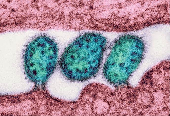
Monoclonal antibodies are among our greatest active in the remedy and prevention of virus-induced diseases. While the highlight focused on Covid-19 monoclonal antibodies during the pandemic, candidate antibodies for other serious pathogens have also progressed. Here we describe a new cocktail of antibodies that neutralize a lesser-known but harmful virus circulating in West Africa: the Lassa virus.
What is Lassa virus?
Lassa virus is a pathogen responsible for Lassa hemorrhagic fever, which affects an additional 300,000 to 500,000 people per year, in West African countries, including Nigeria, Liberia, Sierra Leone, Guinea and Ghana. Lassa fever has a mortality rate of about a year. withcent. Pregnant women are at maximum threat of death, with a mortality rate of up to 90%. Symptomatic cases provide disorders such as fever, headache, vomiting and muscle pain.
Spread occurs regularly through contact with feces or urine from inflamed mice, spread through direct person-to-person contact is also common. SARS-CoV-2, is an RNA virus that constantly mutates. Therapy or prophylaxis is needed to fill the gap in Lassa virus drugs.
Treatment with monoclonal antibodies against Lassa virus
Fortunately, a new candidate could be just around the corner. An organization of researchers at the La Jolla Institute of Immunology in California led by Dr. Anna S. Erica Ollmann Saphire has met a cocktail of 3 antibodies that bind to and neutralize the Lassa virus. This same organization discovered a widely accepted cocktail of monoclonal antibodies against Ebola, which we described above. This cocktail arrived just in time as a regimen to deal with the existing outbreak caused by the Sudanese strain of the Ebola virus in Nigeria.
The new treatment for the Lassa virus, called Arevirumab-3, consists of 3 different antibodies, which bind to different regions of the glycoprotein of the Lassa virus.
The spike protein of the Lassa virus is a set of 3 subunits embedded in the membrane. The virus is synthesized as a long, unmarried polypeptide and cleaved into 3 parts, a main sequence, GP1 encoding the receptor binding function and GP2 incorporated into the membrane (Figure 1).
FIGURE 1: (A) Diagram of LASV GPC design number one showing the positions of NArray-bound subunits and glucans. [ ]. SSP, solid sign peptide; TM, transmembrane domain; tail C, cytoplasmic tail; SKI-1/S1P, subtilisin kexin isoenzyme-1/site-1 protease; Asn, asparagine.
8. 9F inhibits virion cell binding by blocking GPC/α-DG interaction
The first antibody, 8. 9F, binds to the most sensitive tip trimer and spreads over all 3 portions of the glycoprotein. 8. 9F is an antibody because the unpaired antibody binds to all 3 sides of the trimer. The antibody also binds to the 3 cleavage sites of the GP1 subunits. It binds to the glycoprotein component that binds to the host mobile, directly inhibiting the binding of the virion to the mobile by mimicking the mobile alpha-dystroglycan receptor.
The 8. 9F antibody also binds in particular to a glycan, N119, which is to neutralize activity. This glycan is for binding to alpha dystroglycan receptors, so the popularity of 8. 9F is surprising.
A local N89 glycan plays a central role in the recognition of GPC 12. 1F/LASV
The moment the antibody, 12. 1F, binds to a separate site in GP1, it interacts directly with N-glycans to neutralize activity. The antibody binds more to the proximal membrane and has a direct interaction with six amino acid residues and the critical glycans N89 and N109.
To infect a mobile, the Lassa virus will have to bind to the mobile membrane and be engulfed through a mobile endosome. The protein then undergoes structural replacement to bind to the endosome’s internal LAMP-1 protein. The binding of LAMP-1 is essential for the fusion of viral and mobile membranes and access to the cytoplasm where replication occurs.
37. 2D neutralizes LASV by locking the GPC trimer in an inactive configuration.
The third antibody, 37. 2D, binds to two adjacent subunits of GP2. The antibody expands the subunits, blocking the trimer in position through the binding of conserved peptides and conserved glycans N390 and N395. 37. 2D is special because it attacks GP2 at an unmatched angle, stabilizing the prefusion trimer, preventing fusion activation and, consequently, infection.
These 3 mechanisms work in combination to produce a highly effective antibody cocktail.
FIGURE 2: Cryo-EM density maps of local LASV GPC in complex with 8. 9F-scFv/37. 2D-scFv orArray. [ ] Subunits 12. 1F-scFv/37. 2D-scFv. GP1: cyan; GP2 subunits: pink; N-bound glycans: red; TM: grey.
FIGURE 3: (A) Structural style (PDB ID: 7UOV) showing the GPC binding orientation of a 12. 1F-scFv matrix of molecules. [ ]. (B) Epitope of 12. 1F in the GP1 protomer. (C) GP1 link region directed via 12. 1F. Residues are indicated on the interfaces. GP1: blue; 12. 1F HC: green; 12. 1F LC: soft green; the surrounding glycans N89 and N109 are depicted in ball and stick shape. The position of the pH-sensitive region of the histidine triad is also indicated.
A critical question for monoclonal antibody treatment is: Can the virus mutate at binding sites?Dr. Ollmann Saphire’s team shows that this is the case with a single 8. 9 F antibody treatment. However, no resistance mutation appeared when using the cocktail of the three antibodies, nor could the virus escape.
The Arevirumab-3 antibody may provide a long-awaited response to the lack of treatment and prevention of Lassa infections. This is especially important for cases involving pregnant women and their fetuses.
It will be vital to minimize the prices of this antibody cocktail so that it is available when needed in West Africa. Current technologies can produce monoclonal antibodies at $200 and $250 per gram.
The next step in the progression of Lassa fever is the progression of an effective vaccine against all variants of the Lassa virus. This chart can serve as a consultant for the creation of such a vaccine.

