Follow us on social media
Follow us on social media
Follow us on social media
Follow us on social media
Follow us on social media
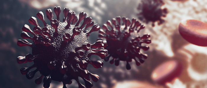
The reaction to the COVID-19 pandemic had to do with action in a multitude of spaces throughout the educational community. We have noticed paintings by engineers to quickly produce effective fanatics, knowledge scientists and epidemiologists who combine to design the possible spread of the virus, economists predict the prosecutor have an effect on pandemics in society, and molecular and cellular biologists run day and night to perceive the nature of the disease and the mechanism of SARS-CoV-2 infection.
In the latter category, various techniques were used, one of which has been shown to be important in the construction of tissues that can become inflamed and broken through the virus, is the use of organoids as models for infection studies.
How can the intestine be affected through COVID-19?
One of the first of these key studies was designed to explore widespread reports of patients with COVID-19 with gastrointestinal disorders in addition to the respiratory disorders that characterize the disease [1].
A collaboration of studies from the Netherlands, led through Mart Lamers (Erasmus Medical Center, Rotterdam, Netherlands) and armed with the wisdom that the virus infects the epithelium of the lungs by interacting with the ACE2 mobile surface receptor, has set out to explore the cause. . of those gastrointestinal symptoms.
Lamers and their team established 3-d moving cultures that had the same spectrum of mobile phones that make up the tissues of the small intestine epithelium. These intestinal organoids were grown under a variety of conditions, leading to disparate numbers of ACE2 receptors in the organoid.
By exposing these organoids to SARS-CoV-2 and tracking the next progression of their electronic infection microscopy, the team was able to identify that the virus was able to infect the intestinal enterocytes of the organoids in their parent and in their mature state.
Enerocyte cells, absorbent cells that shape the epithelium of the small intestine, have been found to produce infectious viral particles, indicating that cells can become inflamed with SARS-CoV-2 and cannot prevent the virus from replicating. The organoid did not vary depending on the proportion of ACE2 receptors present, suggesting that only a very small number of receptors are needed to allow infection.
What brain cells are inflamed with SARS-CoV-2?
As the pandemic continued and reports of new symptoms did not subside, it was obvious that brain tissue needed to be evaluated for possible SARS-CoV-2 infection. Descriptions of headaches, anosmia, confusion, seizures and encephalopathy that accompany the multitude of other COVID symptoms. -19 led a study team from the Chinese Academy of Sciences (Shenzen, China) and the University of Hong Kong (HK) to investigate [2] Array
The team decided to examine a collection of other mobile models, adding human neural progenitor mobiles (hNPCs), neurospheres and brain organoids derived from induced pluripotent mother mobiles.
Brain organoids grew to 35 days, when the presence of fluid-filled ventricular structures was detected, reminiscent of an early cerebral cortex, panneurons, early markers of the anterior brain, and hNPC.
HNCCs were first evaluated to identify their content in ACE2 and other proteins related to coronavirus entry, revealing the presence of ACE2 and a number of other proteins that can allow coronaviruses to enter a cell, building a transparent mechanism through which those cells may be inflamed with SARS-CoV-2.
HNPCs, neurospheres and then brain organoids were challenged with SARS-CoV-2 and SARS-CoV as control. The decision was made that hNPCs were vulnerable to SARS-CoV-2 infection but not SARS-CoV, while brain neurospheres and organoids showed superior protein expression and infectious viral particles, indicating that the virus could infect a human brain.
Other observations of brain organoids revealed that the infection was limited to the positive cells TUJ1- (neural marker) and NESTIN- (NPC marker). This indicates that cortical neurons and HNPC have a maximum threat of infection and suggests that patients who have evolved neurons Symptoms of the disease should be monitored to detect the long-term neurological effects of the disease.
Exploring how COVID-19 causes pneumonia
Organoid lung models that existed prior to the COVID-19 outbreak were temporarily used through organizations to consult their studies and disease, which allowed us to better perceive the virus, however, many organoid models have an imperfect representation of the lungs, limiting the intensity of the data that can be obtained, adding the pathogenic mechanism through which SARS-CoV-2 leads to pneumonia and acute respiratory misery syndrome [3].
A key defect of these models is the result of their inability to maintain the survival and expansion of stem cells that differ in secreting club (AT1) and alveole cells (AT2), either guilty of repairing broken lung tissue. these cells, the “food” cells are used for their expansion, impacting the composition of the organoid making it less representative and therefore less useful.
However, in an article (which has not yet been peer reviewed) on bioRxiv, a collaboration of studies led through Stanford University (California, USA)U. S. ) That included researchers from 10x Genomics (California, USA)U. S. ) And the University of North Carolina at Chapel Hill. (NC, USA) Reports on the success of organoids containing self-innovative AT2 cells capable of transdifferentiated into AT1 cells. These organoids were called AT2 organoids.
They also established “basal organoids” that shaped hairy lighting devices covered with differentiated club mobiles and contained two subsets of basal mobiles containing key mSRa markers of pulmonary basal mobiles. The first set of basal mobiles showed signs of differentiation and proliferation, while the moment showed markers of vesicular and endoplastic reticle metabolism and scaly mobile expression.
This progress already means that organoids provide a much more representative style of the human lung; however, not content with these improvements, the team sought to succeed over the challenge in lung organoids. Extramobileular matrix: the medium through which the organoid would be exposed to SARS-CoV-2. organoids with an eversion approach, expanding the availability of ACE2 luminal receptors for SARS-CoV-2.
Once their organoid models were improved, the team inflamed the organoids with influenza or SARS-CoV-2. They found that the cells were gently inflamed with SARS-CoV-2 and that 72 hours after infection, the viral genome effectively replicated in the cells. along the nucleocapsid protein. 10% of baseline and AT2 cells were inflamed, most of the infection in basal organoids located in AT1 cells.
Obviously, this shows that AT1 and AT2 cells are a new target for SARS-CoV-2 and suggests that their disruption and the next loss of secretory homes would possibly explain, in part, the pathogenesis of pneumonia.
Since the beginning of the pandemic, organoids have allowed researchers to design potentially vulnerable tissues and map infection sites that can contribute to the nebula list of symptoms related to SARS-CoV-2 infection. These studies have known much more than the organs covered in this article as at risk of infection and injury, such as the heart, CNS, and kidneys, but as organoids continue to evolve, driven by the speed of COVID-19 research, they provide much deeper data on the destructive functioning of the disease and the mechanisms of action through which it inflicts its malicious wounds. With this data, the promise of new avenues of protection against the virus arises and the hope of a long term that does not involve accepting COVID-19 as “the new standard”.
BioTechnics is based on Future Science, Future Science Group
44 (0) 20 8371 6090