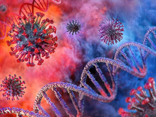
Washington: The headaches, confusion and delirium experienced by some PATIENTS with COVID-19 may be the result of direct invasion of the brain through coronavirus, according to a study published Wednesday.
The studies are still initial, but they provide several new resources of evidence of what was largely unproven in the past.
According to the paper, led by Yale immunologist Akiko Iwasaki, the virus is able to replicate internally in the brain and its presence deprives nearby brain cells of oxygen, the prevalence of this is not yet clear.
Andrew Josephson, director of the neurology branch at the University of California, San Francisco, praised the techniques used in the study and said that “understanding whether there is direct viral involvement in the brain is extremely important. “
But he added that he will remain cautious until the document is peer-reviewed.
It would not be absolutely surprising if SARS-CoV-2 crosses the blood-brain barrier, a pattern that surrounds blood vessels in the brain and attempts to block foreign substances.
Zika virus, for example, also does: it damages the brains of fetuses.
But doctors until now believed that the neurological effects noticed in about one part of all patients could be the result of an immune reaction known as cytokine typhoon that causes inflammation in the brain, rather than direct invasion of the virus.
Iwasaki and his colleagues manipulated the factor in three ways: infecting mini-lab brains called brain organoids, infecting mice, and examining the brain factor from deceased Covid-19 patients.
In brain organoids, the team discovered that the SARS-CoV-2 virus is capable of infecting neurons and then bypassing the machinery of neural cells to make copies of itself.
Inflamed cells promote the death of surrounding cells by disrupting their oxygen supply.
One of the main arguments against the theory of direct invasion of the brain is that the brain lacks the maximum levels of a protein called ACE2 in which the coronavirus is blocked and that is discovered in abundance in other organs such as the lungs.
But the team found that organoids had enough ACE2 to facilitate access to the virus, and proteins were provided in the brain tissue of patients who died.
They also performed a lumbar puncture on a hospitalized Covid-19 patient suffering from delirium and found that the individual had neutralizing antibodies opposed to the virus in his cerebrospinal fluid, additional evidence in favor of their theory.
The team then observed two teams of mice: one that had been genetically modified to have ACE2 receptors in his lungs and the other in his brain.
Other people infected in his lungs showed symptoms of lung damage, while inflammations in the brain temporarily lost weight and died temporarily, indicating potentially greater lethality when the virus enters the organ.
Finally, they tested the brains of 3 patients who died of severe COVID-19 headaches, locating the virus in all of them to varying degrees.
Surprisingly, the inflamed regions have not shown symptoms of infiltration through immune cells, such as T cells, which are quick to attack other viruses such as Zika or herpes to kill the inflamed cells.
This may also simply recommend that the overloaded immune reaction known as cytokine storm, which is guilty of much of the damage that is perceived in the lungs of COVID-19 patients, may not be the main cause of neurological symptoms.
The hypothesis was raised that the nose could simply provide the trace to the brain, but the authors wrote that this should be validated by an additional study.
They added that additional autopsies will be needed to determine the prevalence of brain infection.
Dear reader,
This segment is about life in the United Arab Emirates and data you cannot live without.
Sign up to read and complete gulfnews. com