Researchers from Tsinghua University in Beijing offered new data on the evolution and inter-specific transmission of severe acute respiratory syndrome coronavirus 2 (SARS-CoV-2), the culprit agent of the existing 2019 coronavirus disease pandemic (COVID-19).
Team research into complex viral proteins discovered in SARS-CoV-2 and two very similar coronaviruses revealed differences in their ability to bind and infect host cells that may be the reason SARS-CoV-2 developed such a high infection capacity.
The team learned of significant residues in the complex receptor binding spaces (RBDs) of SARS-CoV-2, Bat Coronavirus RaTG13 and pangolin coronavirus PCoV_GX underlying differences in the activities of those complex proteins and their ability to bind and infect host cells.
Xinquan Wang and his colleagues recommend that five residues, in particular, be related to the evolution of SARS-CoV-2 peak RBD, due to the role they play in the close binding to the conversion of enzyme 2 from the human host angiotensin mobile receptor (hACE2).
“These effects together imply that a strong RBD-ACE2 link and effective RBD shaping sampling are for the evolution of SARS-CoV-2 to download a highly effective infection,” the team wrote.
You must have a preprinted edition of the article in the bioRxiv service, while the article is peer-reviewed.
From animals to humans (zoonotic transmission), coronaviruses pose a risk to human fitness worldwide, as evidenced by the onset of SARS-CoV-1, Middle East Respiratory Syndrome coronavirus (MERS-CoV) and SARS-CoV-2 for more than two decades. .
Current evidence suggests that, like SARS-CoV-1 and MERS-CoV, SARS-CoV-2 is likely local to bats before moving to intermediate hosts and then jumping to humans.
The raTG13 coronavirus, which was detected in horseshoes, known to share the highest significant serial identity (96. 2%) SARS-CoV-2, indicating the maximum likely origin of SARS-CoV-2 in bats.
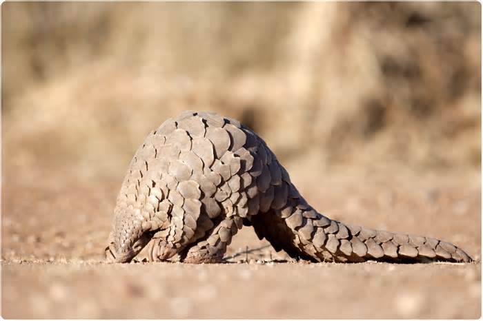
Another Malay pangolin coronavirus (PCoV) known in Guangxi (GX) in China is also very similar to SARS-CoV-2. Genome sequencing of this virus, PCoV_GX, also indicated a higher shared serial identity point (85. 5%) SARS-CoV -2.
As the number one viral design that allows coronaviruses to infect host cells, the role of complex trimer protein in interspecific transmission and infection is of primary interest to researchers.
“Complex coronavirus glucoproteins recognize their mobile host receptor and mediate membrane fusion for entry, acting as well as the ultimate critical coronavirus protein for viral evolution and interspeciific transmission,” Wang and his colleagues explain.
Cryogenic electron microscopy (cryo-EM) studies have shown in the past that, like the SARS-CoV-1 complex trimer, the SARS-CoV-2 complex trimer should have at least one RBD in an “upward” conformation to bind hACE2.
“Therefore, a peak trimer with the 3 RBD” below “is in an inactive receptor binding state, and the conformal replacement of at least one RBD from” under “up” causes the complex trimer to move to an active binding state to the receptor, the researchers explain.
Now, Wang and his colleagues have made a decision on the cryo-EM structures of complex proteins from the RaTG13 and PCoV_GX peaks and compared to the SARS-CoV-2 peak.
The research revealed that RaTG13 and PCoV_GX RBDs looked a lot like those of the SARS-CoV-2 peak.
The 3 RBDs of the raTG13 and PCoV_GX complex trimers were in “downward” shaping, suggesting that those RBDs have a tendency to adopt the inactive state of binding to the receiver.
However, by conducting surface plasmonic resonance experiments, the researchers discovered that the PCoV_GX RBD peak had a binding affinity for hACE2 to that of the SRAS-CoV-2 RBD peak. At the same time, the RaTG13 RBD demonstrated a much weaker hACE2 link.
The team then learned diversifications at the point of six residues in the RBD that gave the impression of these differences in the binding of hACE2 between the 3 viruses.
The Y449, Q493, Q498, N501, and Y505 relays were regrouped to form a patch on the SARS-CoV-2 RBD that interacted strongly with hACE2.
Researchers also learned about two-position amino acid adjustments (Y449 and Y505) that occurred only at the RaTG13 RBD peak and not at the PCoV_GX RBD peak, which researchers believe may simply decrease the hACE2 binding of raTG13.
“We further propose that the patch containing Y449, Q493, Q498, N501 and Y505 plays a critical role in the evolution of SARS-CoV-2 RBD, selling a close link in hACE2 and impacting the other affinities observed between RBD and ACE2 Orthologists in wild and domestic animals,” Wang and his colleagues write.
The team also met 3 N-linked glycosylations in raTG13 and the RBD peak of PCoV_GX, one of which (N370) is not a glycosylation at the RBD peak of SARS-CoV-2.
“The absence of N370-related glucans would possibly contribute to greater flexibility in SARS-CoV-2 pic RBDs,” the researchers suggest.
They say this speculation is supported by some other study that appears that the N165 mutation in SARS-CoV-2 resulted in an accumulation in the “upward” conformation of RBDs, suggesting that glucans serve as a conformal detail of RBD.
”Based on all these results, we propose that the RBD-hACE2 close link we observe is the most critical to determining the effectiveness of various cellular inputs between RaTG13, PCoV_GX and SARS-COV-2”, the researchers explained.
“This and the replacement of the RBD conformation” from the bottom up “are for the evolution of SARS-CoV-2 to obtain a very effective transmission capacity”, concludes the equipment.
bioRxiv publishes initial clinical reports that are not peer reviewed and should therefore not be considered conclusive, the consultant’s clinical practice/health-related behaviors, nor treated as established information.
Wang X and cabbage. The glycoprotein structures of bat and pangolin peak coronavirus provide data on the evolution of SARS-CoV-2. bioRxiv, 2020. doi: https://doi. org/10. 1101/2020. 09. 21. 307439
Written by
Sally holds a bachelor’s degree in biomedical sciences (B. Sc. ). He specializes in reviewing and synthesizing the latest discoveries in all covered medical spaces in major world-renowned, high-impact foreign medical journals, foreign press conferences, and newsletters from government agencies and regulators. At News-Medical, Sally generates news, articles about life sciences, and interview coverage.
Use one of the following to cite this article in your essay, job, or report:
Apa
Robertson, Sally. (2020, 23 September). The design of bat and pangolin coronavirus shows the evolution of SARS-CoV-2 News-Medical. Retrieved September 23, 2020 in https://www. news-medical. net/news/20200923/Bat-and -pangolin-coronavirus-layout-sheds-gentle-on-SARS-CoV-2-evolution. aspx.
Mla
Robertson, Sally. ” The design of bat and pangolin coronavirus gently influences the evolution of SARS-CoV-2″ . News-Medical. 23 September 2020.
Chicago
Robertson, Sally. ” The design of bat and pangolin coronavirus gently influences the evolution of SARS-CoV-2″ . News-Medical. https: //www. news-medical. net/news/20200923/Bat-and-pangolin- coronavirus-layout-sheds-gentle-on-SARS-CoV-2-evolution. aspx. (accessed 23 September 2020).
Harvard
Robertson, Sally. 2020. La layout of bat and pangolin coronavirus, sheds gentil on the evolution of SARS-CoV-2. News-Medical, accessed September 23, 2020, https://www. news-medical. net/news/ 20200923 / Sheds-design-de-coronavirus-bat-y-pangolin-soft-over-SARS-CoV-2-evolution. aspxArray
News-Medical. net – An AZoNetwork site
Ownership and operation through AZoNetwork, © 2000-2020

